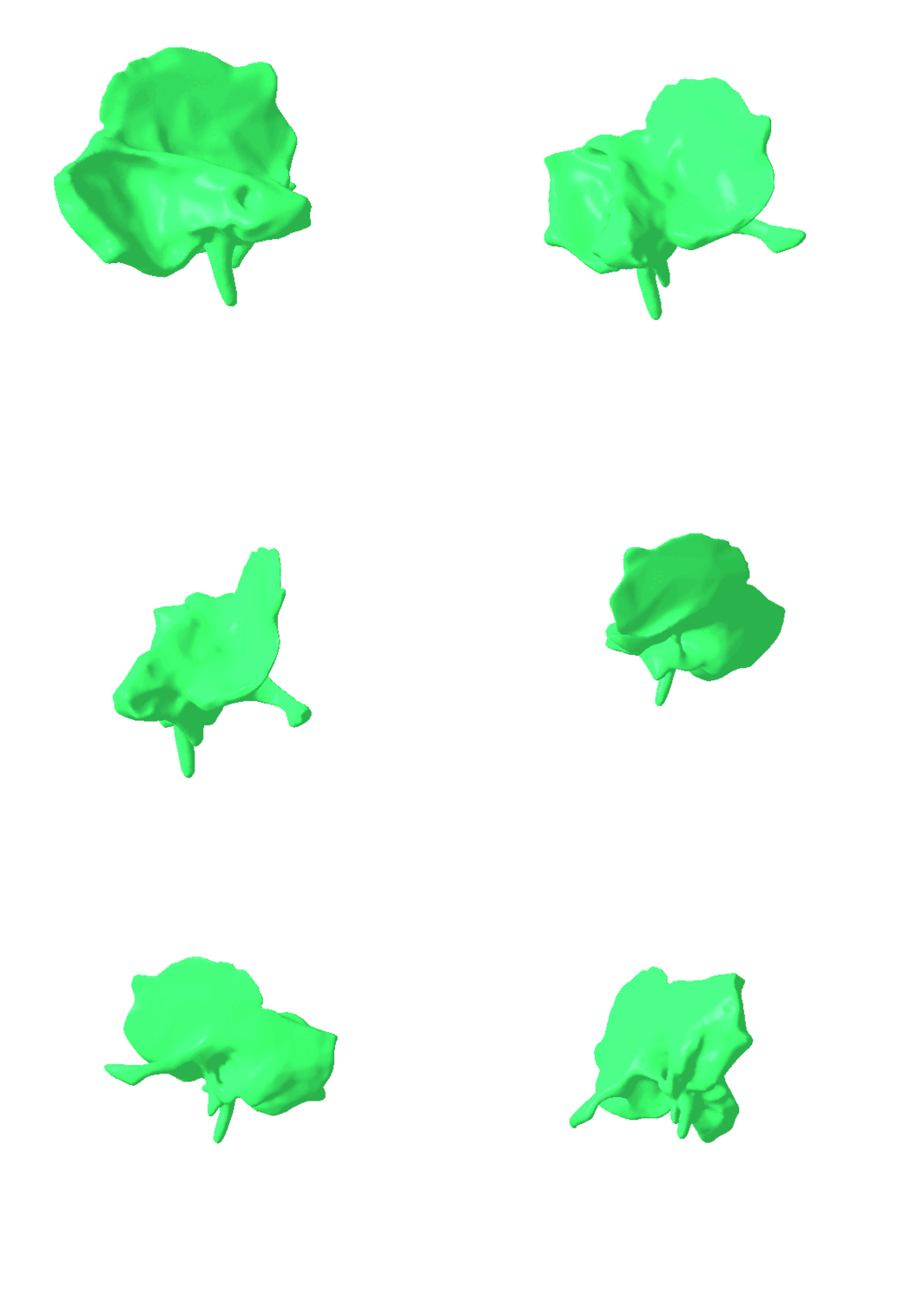
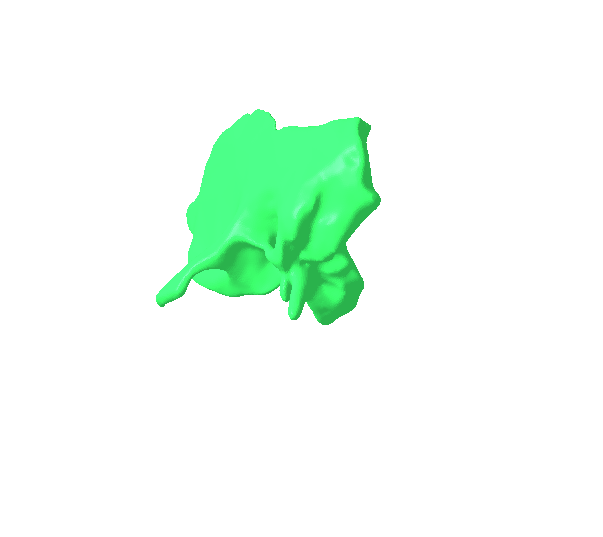
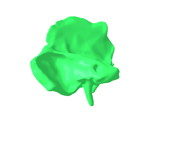
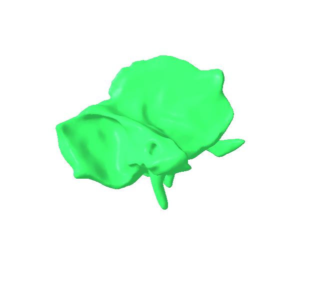
This is high detailed segmented model.
Size 14.3 MB+ 13.5 MB without zip.
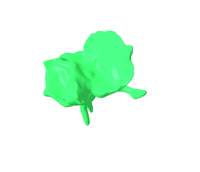
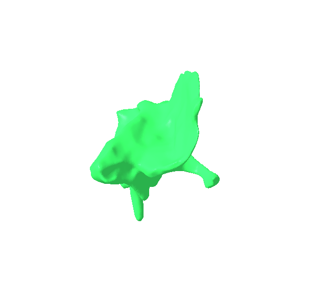
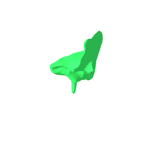
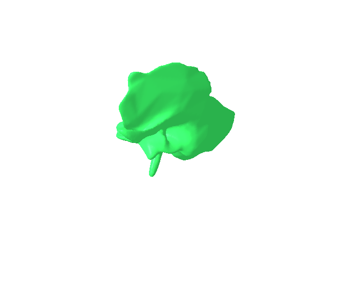
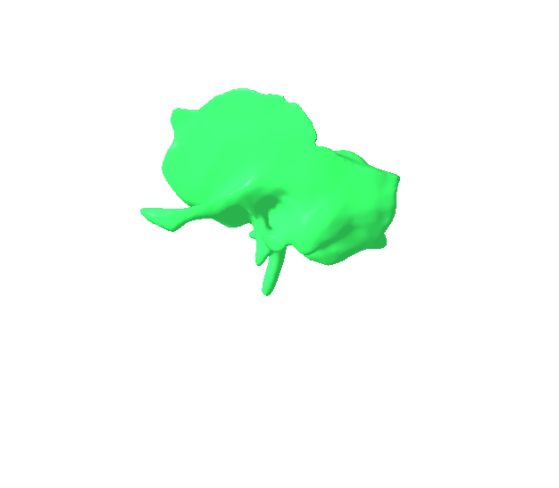
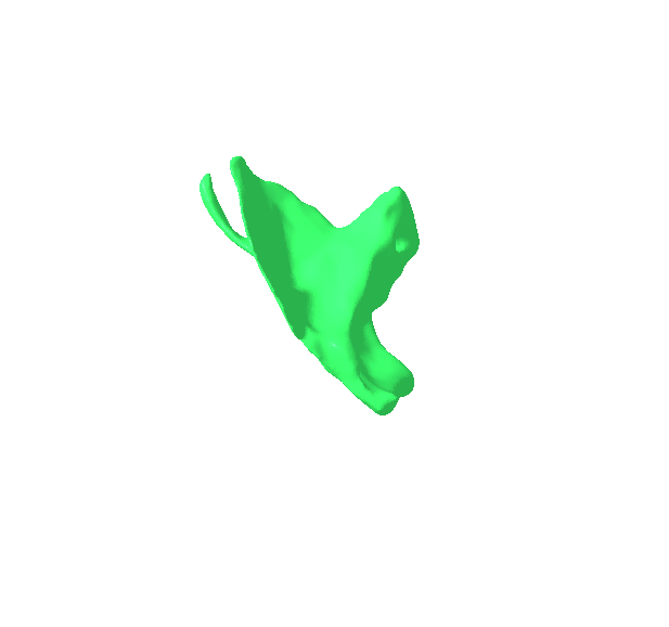
The left temporal bone is one of the two temporal bones of the skull, situated on the left side of the head, forming part of the lateral (side) wall and base of the cranium. As a highly complex and irregular bone, it is indispensable for auditory function, balance, jaw movement, and safeguarding vital neurological and vascular structures.Each temporal bone is uniquely shaped and is conventionally divided into several distinct parts:
Squamous Part:
This is the largest and most superior portion, characterized by its thin, fan-like shape that forms part of the lateral aspect of the skull.
It features the zygomatic process, a slender, forward-projecting bony arch that articulates with the temporal process of the left zygomatic bone, together forming the left zygomatic arch (commonly known as the cheekbone).
On its inferior surface, just anterior to the external ear opening, lies the mandibular fossa (glenoid fossa). This specific depression articulates with the condyle of the left mandible, thereby forming the left temporomandibular joint (TMJ), which is essential for all movements of the lower jaw, including chewing and speaking.
Tympanic Part:
A relatively small, C-shaped bone plate that forms the anterior, inferior, and posterior walls of the external acoustic meatus (the external ear canal), which leads directly to the eardrum (tympanic membrane).
It also contributes to the floor of the ear canal.
Projecting downwards and forwards from its inferior surface is the styloid process, a slender, pointed bony projection. This process serves as a crucial attachment point for several muscles and ligaments associated with the tongue and pharynx (e.g., stylohyoid, styloglossus, stylopharyngeus muscles).
Petrous Part:
Recognized as the hardest and most dense portion of the temporal bone, it is pyramid-shaped and extends medially (inward) into the base of the skull. Its robust nature provides excellent protection.
It encapsulates the middle ear cavity (containing the malleus, incus, and stapes, which are essential for sound conduction) and, more importantly, the intricate inner ear structures. The inner ear comprises the cochlea (the organ responsible for hearing) and the vestibule and semicircular canals (the organs responsible for maintaining balance).
Within the petrous part, the internal acoustic meatus is a vital canal that allows the passage of the facial nerve (CN VII), vestibulocochlear nerve (CN VIII), and associated blood vessels from the brainstem to the inner ear and facial muscles.
This part also contains the carotid canal (a passageway for the internal carotid artery) and contributes to the formation of the jugular foramen (along with the occipital bone), through which the internal jugular vein and cranial nerves IX, X, and XI pass.
Mastoid Part:
Located posterior to the tympanic part and inferior to the squamous part, this portion is characterized by the prominent, palpable mastoid process. This is a conical projection easily felt behind the ear.
The mastoid process is extensively pneumatized, meaning it contains numerous interconnected mastoid air cells that communicate with the middle ear cavity.
It provides a substantial attachment area for several powerful neck muscles, most notably the sternocleidomastoid muscle.
Articulations (Joints):
The left temporal bone articulates with five other bones of the skull:
Left Parietal bone: Superiorly, via the squamosal suture.
Left Occipital bone: Posteriorly, via the occipitomastoid suture.
Left Sphenoid bone: Anteriorly, via the sphenosquamosal suture.
Left Zygomatic bone: Anteriorly, forming the zygomatic arch.
Left Mandible: Inferiorly, forming the temporomandibular joint (TMJ).
Functions:
Hearing and Balance: Its petrous part is the bony encasement for the highly sensitive organs responsible for these vital senses.
Brain Protection: Forms part of the cranial vault, providing protection for the left temporal lobe of the brain.
Jaw Function: Forms the socket component of the left TMJ, enabling precise movements of the lower jaw for chewing, speaking, and other functions.
Muscle Attachment: Serves as an extensive attachment site for various muscles of mastication, facial expression, and the neck, influencing head movement and facial features.
Neurovascular Passageway: Provides critical canals and foramina for the passage of major nerves and blood vessels to and from the brain, face, and neck regions.#LeftTemporalBone #TemporalBone #SkullAnatomy #CranialBones #HumanAnatomy #EarAnatomy #Hearing #Balance #TMJ #TemporomandibularJoint #MandibularFossa #SquamousPart #PetrousPart #TympanicPart #MastoidProcess #ZygomaticArch #StyloidProcess #InnerEar #MiddleEar #ExternalAcousticMeatus #FacialNerve #VestibulocochlearNerve #CarotidCanal #JugularForamen #MastoidAirCells #Neuroanatomy #SkeletalSystem #BoneAnatomy #AnatomyAndPhysiology #MedicalEducation #Otolaryngology #Neurosurgery #HeadAndNeckAnatomy #SkullBase #CranialVault #BoneHealth #HumanBody #BiologicalSystems #Osteology #TemporalLobe #EarHealth #HearingLoss #Vertigo #OtitisMedia #Mastoiditis #TMD #Mandible #Articulations #Sutures #AnatomicalStudy #ClinicalAnatomy



:format(webp)/https://fbi.cults3d.com/uploaders/37864523/illustration-file/6825bd51-4f32-4c74-ad61-e6982ca824c7/Gif.gif)