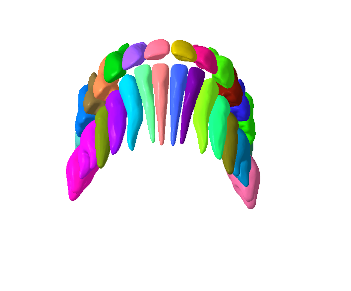
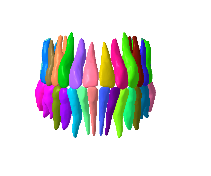
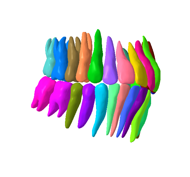
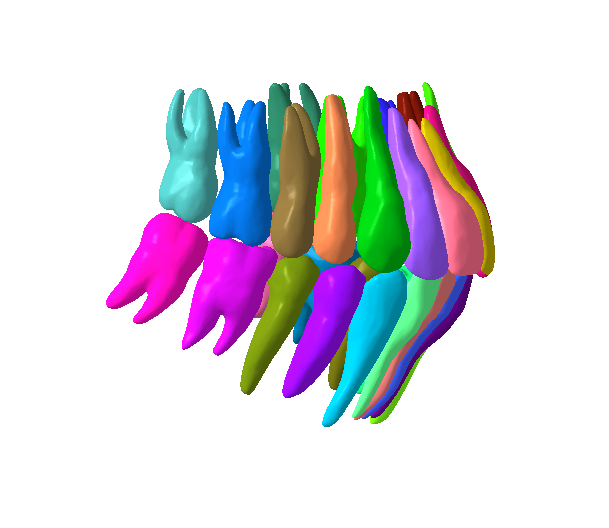
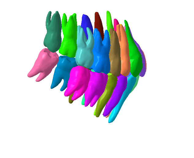
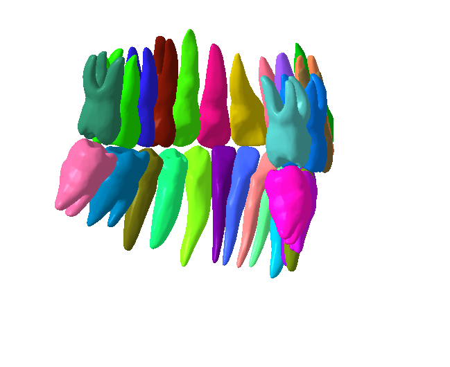
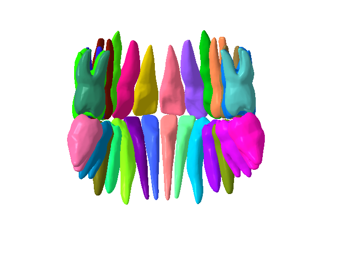
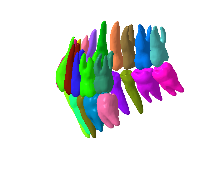
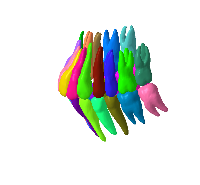
This is high detailed segmeted model.
Size 940 MB without zip. Each teeth around 40 MB.
28 Separate Teeth files.
Human teeth are complex, calcified structures embedded in the jawbones, primarily designed for mastication (chewing food), but also crucial for speech articulation and facial aesthetics. They are the hardest substances in the human body.Humans typically have two sets of teeth during their lifetime:
Deciduous Teeth (Primary/Baby Teeth): There are usually 20 deciduous teeth (10 in the upper jaw, 10 in the lower jaw). They begin to erupt around 6 months of age and are usually all present by 2-3 years. They serve to hold space for the permanent teeth and aid in early chewing and speech development.
Permanent Teeth (Secondary/Adult Teeth): These replace the deciduous teeth, starting around age 6, and a full set usually consists of 32 teeth (16 in the upper jaw, 16 in the lower jaw), including wisdom teeth.
Types of Permanent Teeth:
Each type of tooth is specialized for a particular function:
Incisors (8 total): The four front teeth in both the upper and lower jaws. They are sharp, chisel-shaped, and used for cutting and biting food.
Canines (4 total): Located at the "corners" of the mouth, one in each quadrant. They are pointed and strong, used for tearing food.
Premolars (Bicuspids) (8 total): Located behind the canines. They have two cusps (points) and a flatter surface, used for crushing and grinding food.
Molars (12 total): The largest teeth, located at the back of the mouth. They have broad, flat chewing surfaces with multiple cusps, designed for grinding and pulverizing food before swallowing. The third molars are commonly known as "wisdom teeth."
Structure of a Tooth:
Each tooth has two main parts:
Crown: The visible part of the tooth, projecting above the gum line.
Enamel: The outermost layer, covering the crown. It's the hardest substance in the body, protecting the tooth from decay and wear.
Dentin: Lies beneath the enamel and cementum. It's a bone-like material, less hard than enamel, forming the bulk of the tooth. It contains microscopic tubules that transmit sensations.
Pulp Cavity: The innermost part of the crown, containing the pulp.
Root(s): The part embedded within the jawbone, below the gum line.
Cementum: A thin, bone-like layer covering the root surface. It anchors the tooth to the periodontal ligament.
Pulp Canal (Root Canal): Extends from the pulp cavity through the root(s).
Pulp: Soft tissue within the pulp cavity and root canal, containing blood vessels, nerves, and connective tissue. It provides nourishment and sensation to the tooth.
Supporting Structures (Periodontium):
Teeth are held in place by the periodontium, which includes:
Gingiva (Gums): Soft tissue surrounding the teeth.
Periodontal Ligament (PDL): Fibers that connect the cementum of the tooth root to the alveolar bone, acting as a shock absorber.
Alveolar Bone: The part of the jawbone that holds the tooth sockets.
Functions:
Mastication: Breaking down food into smaller, manageable pieces for digestion.
Speech: Essential for forming certain sounds and clear pronunciation.
Facial Structure and Aesthetics: Maintain the shape of the face, support the lips and cheeks, and contribute significantly to a person's smile and appearance.
#HumanTeeth #Dentition #OralHealth #DentalAnatomy #ToothStructure #Enamel #Dentin #Pulp #Gingiva #Gums #Periodontics #DentalCare #OralHygiene #HealthySmile #Mastication #Incisors #Canines #Premolars #Molars #WisdomTeeth #DeciduousTeeth #PermanentTeeth #OralCavity #Jawbone #AlveolarBone #PeriodontalLigament #ToothDecay #Cavities #DentalFillings #RootCanal #ToothExtraction #Orthodontics #Braces #DentalImplants #Crowns #Veneers #Dentistry #DentalFacts #OralWellness #DentalClinic #SmileTransformation #OralSurgery #Endodontics #Prosthodontics #PreventiveDentistry #DentalEducation #OralHealthMatters #ToothDevelopment #FacialAesthetics


:format(webp)/https://fbi.cults3d.com/uploaders/37864523/illustration-file/6825bd51-4f32-4c74-ad61-e6982ca824c7/Gif.gif)
:still()/https://fbi.cults3d.com/uploaders/37864523/illustration-file/44486aad-9968-409d-851b-3cff5ac8e25a/8.gif)
:still()/https://fbi.cults3d.com/uploaders/37864523/illustration-file/100e6cc3-b36f-40cb-a16f-bd650e48c2dd/10.gif)
:still()/https://fbi.cults3d.com/uploaders/37864523/illustration-file/080ea5c9-e3f6-4fb0-895c-56b95c586b98/10.gif)
:still()/https://fbi.cults3d.com/uploaders/37864523/illustration-file/da1e49ef-22ea-4a2e-88e3-1359647be421/10-2.gif)
:still()/https://fbi.cults3d.com/uploaders/37864523/illustration-file/a5ee9cd5-75a3-4e7f-bd5a-accf719cb9ee/10.gif)
:still()/https://fbi.cults3d.com/uploaders/37864523/illustration-file/4e12e323-5467-43ef-a322-e20f06f19c37/12.gif)
:still()/https://fbi.cults3d.com/uploaders/37864523/illustration-file/c89aff48-d0b2-439c-b962-eb6de6863f7d/13.gif)