This is high detailed model.
Size - 32 MB without zip.
1 file.
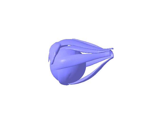
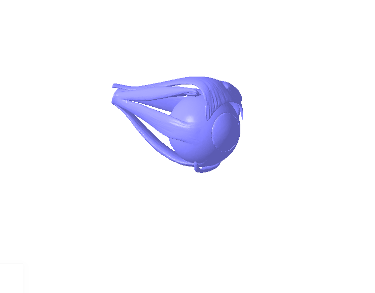
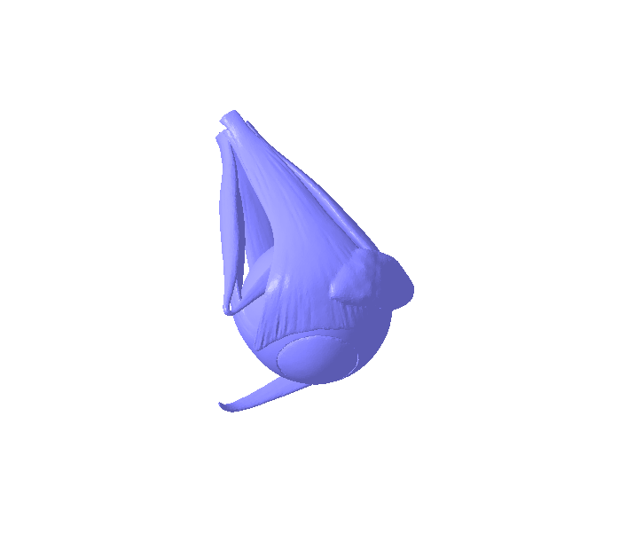
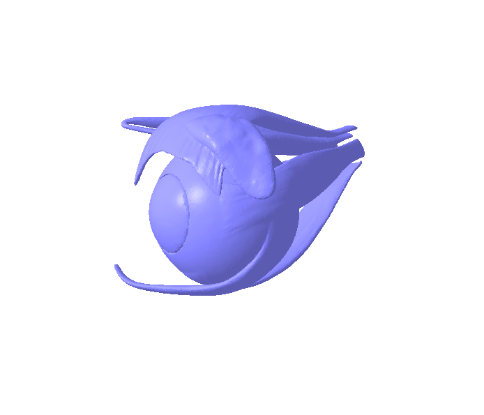
The orbit (specifically, the left orbit in this case) is a roughly pyramidal bony cavity in the skull, with its base facing forward and its apex pointing posteriorly (towards the back of the head). It is formed by the articulation of seven cranial and facial bones:
Frontal bone: Forms the superior (upper) wall and part of the medial wall.
Zygomatic bone: Forms the lateral (outer) wall and part of the floor.
Maxillary bone: Forms most of the floor and part of the medial wall.
Ethmoid bone: Forms a significant portion of the medial wall.
Lacrimal bone: Forms part of the medial wall, anterior to the ethmoid.
Palatine bone: Contributes a small part to the posterior floor.
Sphenoid bone: Forms parts of the lateral and posterior walls (including the greater and lesser wings) and the apex of the orbit, housing important foramina (openings).
Key Contents within the Left Orbit:
Beyond the bony walls, the orbital cavity contains a multitude of essential structures that support the function of the left eye:
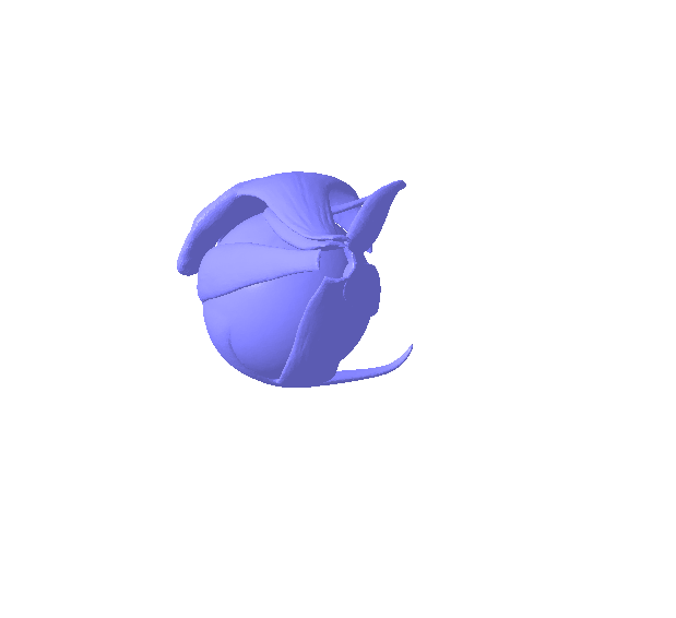
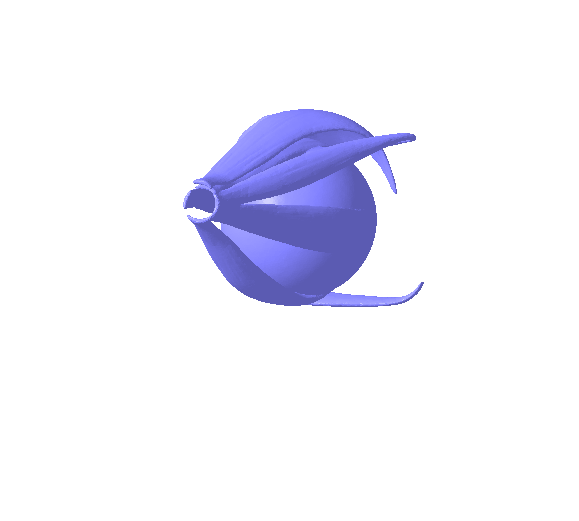
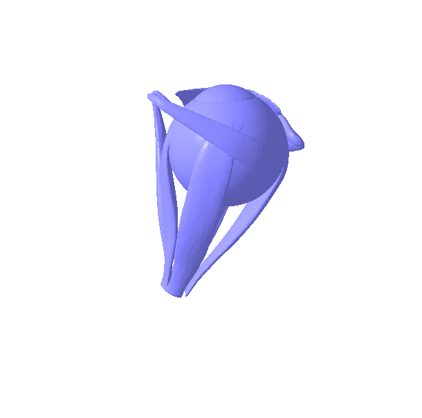
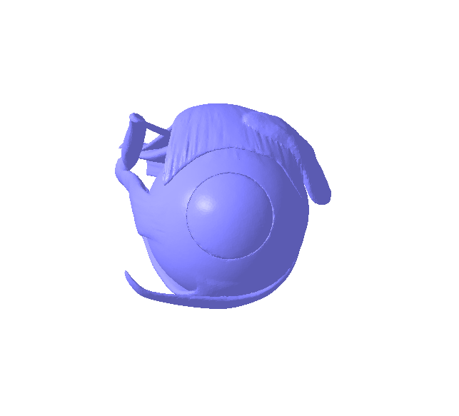
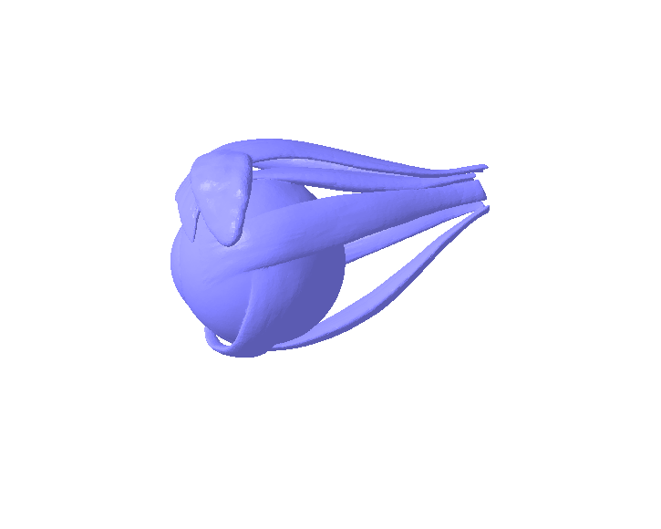
Left Eyeball: The primary occupant, responsible for vision.
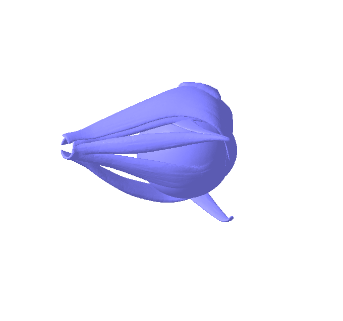
Extraocular Muscles: Six extrinsic muscles (Superior Rectus, Inferior Rectus, Medial Rectus, Lateral Rectus, Superior Oblique, Inferior Oblique) that attach to the eyeball and control its precise movements, allowing gaze direction.
Optic Nerve (Cranial Nerve II): Transmits visual information from the retina of the eye to the brain. It exits the orbit through the optic canal in the sphenoid bone.
Oculomotor Nerve (Cranial Nerve III): Primarily supplies most of the extraocular muscles.
Trochlear Nerve (Cranial Nerve IV): Supplies the superior oblique muscle.
Abducens Nerve (Cranial Nerve VI): Supplies the lateral rectus muscle.
Trigeminal Nerve Branches (e.g., Ophthalmic division - CN V1): Provide sensory innervation to the eye, orbit, and surrounding facial areas.
Ophthalmic Artery: The main blood supply to the orbital structures and eyeball, branching from the internal carotid artery.
Ophthalmic Veins: Drain blood from the orbital structures.
Lacrimal Gland: Located in the upper outer part of the orbit, it produces tears to lubricate and protect the eye.
Orbital Fat: Acts as a protective cushion for the eyeball and other structures, allowing smooth movement.
Ligaments and Fascia: Connective tissues that support and suspend the contents within the orbit.
Functions of the Left Orbital Part:
Protection: The bony walls provide robust protection against external trauma to the delicate eyeball and its associated structures.
Support: It provides a stable base and attachment points for the extraocular muscles, allowing precise eye movements.
Passageway: Contains various foramina and fissures (e.g., superior orbital fissure, inferior orbital fissure, optic canal) that allow the passage of nerves, arteries, and veins to and from the cranial cavity and other facial regions.#LeftOrbit #EyeSocket #OrbitalAnatomy #HumanEye #Eyeball #CranialBones #FacialBones #Ophthalmology #Neuroanatomy #EyeProtection #ExtraocularMuscles #OpticNerve #OculomotorNerve #TrochlearNerve #AbducensNerve #OphthalmicArtery #OrbitalFat #LacrimalGland #OrbitalFracture #SkullAnatomy #AnatomyAndPhysiology #VisionScience #OcularHealth #EyeCare #SurgicalAnatomy #OrbitSurgery #MedicalImaging #CTScan #MRI #EyeMovement #VisualSystem #HeadAndNeckAnatomy #BonyOrbit #OrbitalContents #EyeHealth #OrbitalDisease #Proptosis #Enophthalmos #SkullBase #SphenoidBone #FrontalBone #ZygomaticBone #MaxillaryBone #EthmoidBone #LacrimalBone #PalatineBone #Foramina #OrbitalFissure #AnatomicalStudy #ClinicalOphthalmology


:format(webp)/https://fbi.cults3d.com/uploaders/37864523/illustration-file/6825bd51-4f32-4c74-ad61-e6982ca824c7/Gif.gif)